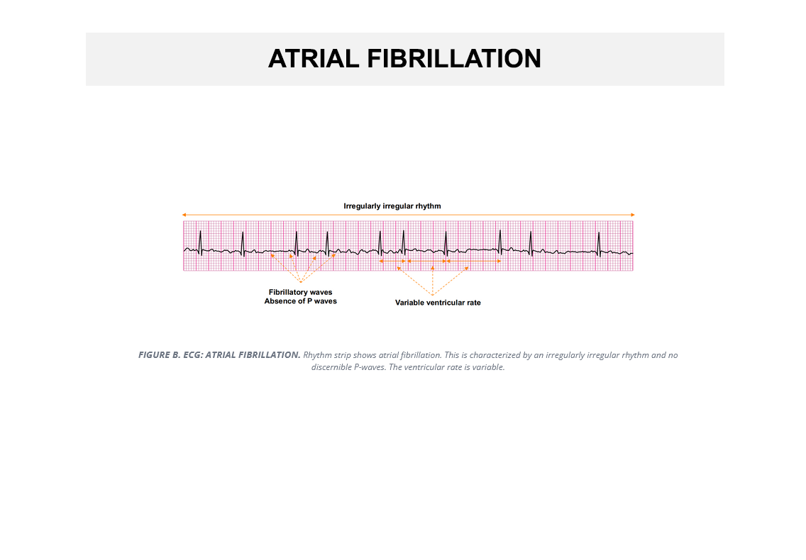
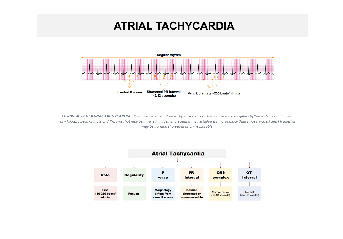
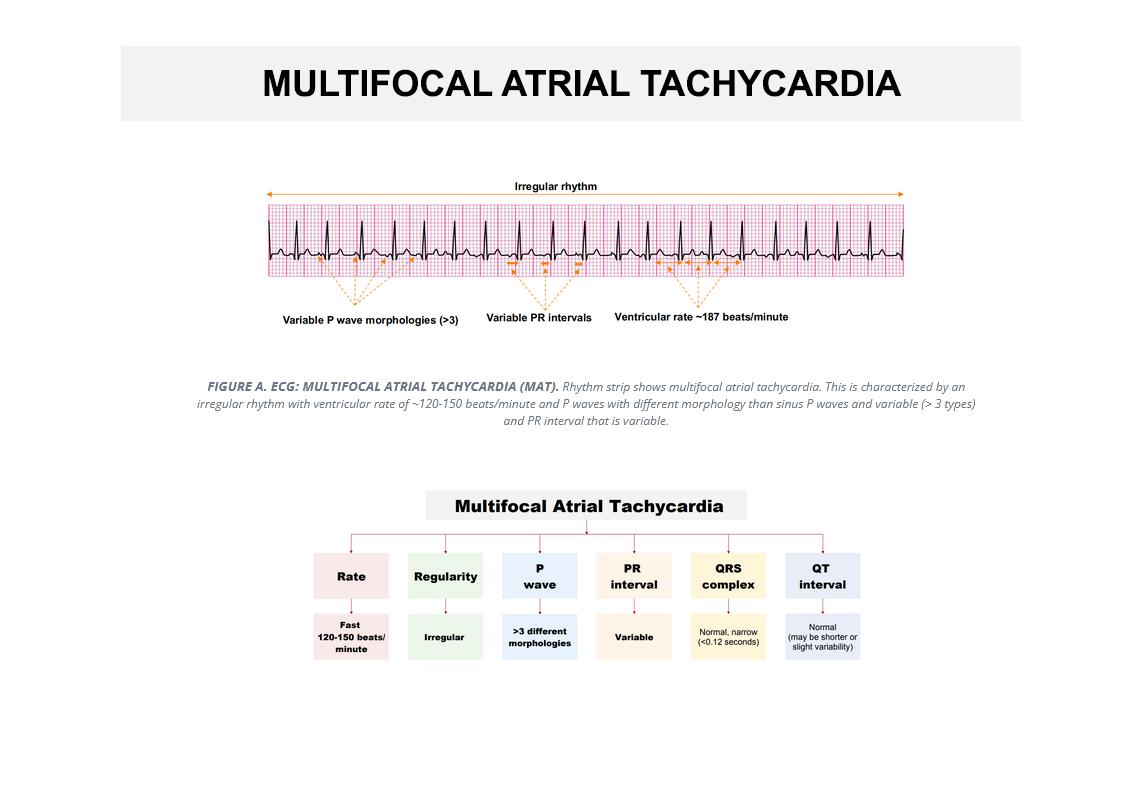
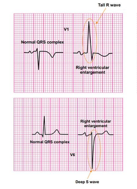
Systematic Approach to EKG and Interpretation for PANCE

Systematic EKG Interpretation for PANCE

This material provides comprehensive guidance on anatomy and physiology, the use of a heart monitor, and the skills needed to identify and interpret EKG tracings, including key patterns and abnormalities.

  • Identifying normal sinus rhythms
  • Cardiac and respiratory syndromes
  • Arrhythmias: sinus, atrial, junctional, and ventricular
  • AV conduction issues
  • Pacemaker and ICD management
  • Medication side effects
  • Bundle branch block detection
  • 3-lead, 5-lead, and 12-lead EKG recordings
  • Detailed electrode placement illustrations
  • Electrolyte imbalances and metabolic influences
  • Protocols for routine diagnostic tests

Study abnormal EKG tracings
for PANCE

Reviewing Abnormal EKG Tracings
for PANCE

This EKG review offers a systematic approach to rhythm interpretation, aiding in the diagnosis, monitoring, and assessment of the severity of conditions such as:

  • Inherited heart conditions
  • Blockages in coronary arteries
  • Acute myocardial infarction (Heart attack)
  • Heart muscle weakness (Heart failure)
  • Abnormal heart sounds (Murmurs)
  • Heart muscle disorders (Cardiomyopathy)
  • Inflammation of the heart's outer layer (Pericarditis)
  • Irregular heartbeats (Arrhythmias)

Study EKG patterns and cardiac structural abnormalities for PANCE

Improving EKG Interpretation and Recognizing Structural Issues for PANCE

Improve your ability to accurately interpret EKG tracings to spot irregularities and abnormalities, a core competency for the PANCE exam.

Learn to recognize the effects of medications and drug use, such as amphetamines, over-the-counter cold and allergy medications, beta blockers, and cocaine, on the heart’s electrical system.

Understand how to identify structural heart abnormalities, like chamber or wall enlargement, that could signal potential heart problems or defects.

Review EKG Conduction Abnormalities for the PANCE

Streamline EKG Interpretation for the PANCE with an Organized Approach

Classification of supraventricular tachycardia

Visually rich Qbank graphics

More than 3,000 highly detailed graphics, tables, diagrams, flow charts and illustrations.

The UMock methodology helps organize the underlying basic concepts with a high quality presentation of visual aids.

The use of visual aids from UMock makes it easier to understand complex concepts, improve retention, and reduce study time.

Complete Pance Online Course Review
Choose The Best Plan

EKG Course Review

Course Content

Expand All

● Cardiac Muscle

● Cardiac Chambers

● Coronary Artery Anatomy

● Cardiac Conduction System

● Cardiac Action Potentials

● Cardiac Innervation

● Cardiac Cycle

● Electrocardiogram

● ECG Electrodes

● Limb Leads

● Precordial Leads

● Contiguous Leads

● ECG Paper

● ECG Recording

● ECG Waves

● ECG Segments and Intervals

● Heart Rate Determination

● Cardiac Axis Determination

● Electrode Misplacement

● P Wave

● PR Interval

● QRS Complex

● R Wave Progression

● ST Segment

● T Wave

● QT Interval

● U Wave

● Atrial Enlargement

● Ventricular Enlargement

● Classification of Arrhythmias

● Tachyarrhythmias

● AV Nodal Reentry Tachycardia (AVNRT)

● Classification of AVNRT

● Atrioventricular Reentry Tachycardias (AVRT)

● Triggered Activity

● Wide QRS Tachyarrhythmias

● VT versus SVT

● Vereckei Algorithm

● Brugada Algorithm

● Adenosine

● Adenosine Effect on Arrhythmias

● Sinus Tachycardia

● Sinus Bradycardia

● Sinus Dysrhythmias

● Sinus Arrest and Pause

● Sick Sinus Syndrome

● Atrial Arrhythmias

● Wandering Atrial Pacemaker

● Premature Atrial Complexes

● First Degree Heart Block

● Second Degree Heart Block

● Third Degree Heart Block

● Atrial Flutter

● Atrial Fibrillation

● Atrial Tachycardia

● Multifocal Atrial Tachycardia

● Premature Junctional Beat

● Early Repolarization

● Wolf-Parkinson-White Syndrome

● Junctional Tachycardia

● Benign Early Repolarization

● Ventricular Arrhythmias

● Premature Ventricular Complexes

● Couplets and Bigeminy

● Trigeminy

● Idioventricular Rhythm

● Ventricular Tachycardia

● Torsade de Pointes

● Ventricular Fibrillation

● Asystole

● Hyperkalemia

● Hypokalemia

● Hypercalcemia

● Hypocalcemia

● Hyperthyroidism

● Hypothyroidism

● Digitalis Effect

● ß-Blocker Toxicity

● Sotalol Toxicity

● Calcium Channel Blockers Toxicity

● Quetiapine Toxicity

● Electrical Alternans

● Pericarditis

● Pulmonary Embolism

● Angina

● Acute Coronary Syndrome

● Myocardial Infarction

● STEMI

● Sudden Cardiac Death

● Hypertrophic Cardiomyopathy

● Cardiac Muscle

● Cardiac Action Potential

● WolfParkinson-White Syndrome

● Hypothermia

● Wellen’s Syndrome

● Myocardial Infarction

● ACS Syndrome

● STEMI

● Pulmonary Embolism

● Brainstem Herniation

● De Winter’s ECG Sign

● Sotalol Toxicity

● Propranolol Toxicity

● Tricyclic Antidepressant Toxicity

EKG Book Content

PANCE EKG Interpretation

Prepare for your exam with UMock EKG Review. Each chapter includes a
complete review of topics with a Q&A at the end of the book.

Sinus Rhythms
Learn how to identify features and qualifying criteria of sinus rhythms including normal, sinus bradycardia, sinus tachycardia, sinus dysrhythmia, sinus arrest, and sinus exit block.
Atrial Rhythms
Recognize complexes and rhythms for premature atrial complexes, wandering atrial pacemaker rhythm, multifocal atrial tachycardia, atrial flutter, and atrial fibrillation.
Junctional Rhythms
Identify features and criteria for premature junctional complexes, junctional escape beats, accelerated junctional rhythm, junctional tachycardia, and supraventricular tachycardia.
Heart Block Rhythms
Review the features and criteria for first degree heart block, second degree heart block type I, second degree heart block type II, and third degree/complete heart block.
Ventricular Rhythms
Identify features of ventricular rhythms including premature ventricular complexes, agonal rhythm, idioventricular and accelerated idioventricular rhythm, ventricular tachycardia, ventricular fibrillation, and asystole.
Pacemaker Rhythms
Learn how to recognize features of pacemaker rhythms including atrial and ventricular pacemaker rhythm, atrioventricular pacemaker rhythm, and failure to capture.
12-Lead ECG Axis
Review of which major coronary artery supports each area of the heart and learn the fundamentals of electrical axis determination and the presence of cardiac axis deviation.
Performing 12-Lead
Learn 12-lead EKG tracings which presents the electrical activity of the heart on two planes. Leads I, II, III, aVR, aVL, and aVF give a flat view of the heart from surrounding edges of a frontal plane.
Bundle Branch Block
Recognize the features for the presence of a complete left or right bundle branch block.
Left Ventricular Hypertrophy
Learn how to recall and apply the process for measurement to determine the presence of left ventricular hypertrophy.
Injury, Ischemia & Infarction
Review of the fundamentals and differences of indicative and reciprocal views, and recognize EKG changes caused by STEMI, ischemia, injury, and infarction.
EKG Terminology
Learn basic EKG terminology including PR interval, QRS complex, QT interval and ST interval.

" The EKG review gave me a complete understanding of accurately interpreting tracings for various heart conditions. The content covers everything you’ll see on PANCE related to analyzing and interpreting EKG tracings.
This is the only source you’ll need for learning EKG for the PANCE exam. It’s a complete guide with plenty of step by step procedural information that covers the entire spectrum of abnormalities you’ll need to recognize on EKG tracings. "

Hailey

New York University

0